
By request
by James Lyons-Weiler, PhD, Popular Rationalism, ©2026
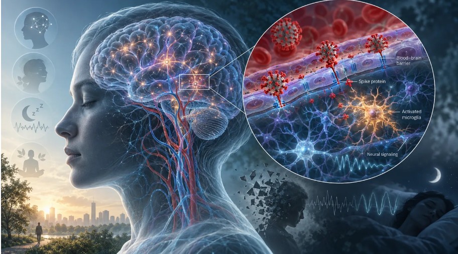
(Sep. 27, 2026) — The relationship between SARS-CoV-2 infection and mental health has often been framed too narrowly. One interpretation attributes depression, anxiety, cognitive dysfunction, sleep disruption, fatigue, and related complaints largely to the psychological burden of illness, isolation, social disruption, and pandemic stress. Another attributes these outcomes directly to the biological effects of the virus, and increasingly to the SARS-CoV-2 spike protein itself.
Neither formulation adequately represents the evidence.
Human studies have established that neurological, cognitive, and psychiatric abnormalities can follow SARS-CoV-2 infection. Experimental studies have separately demonstrated that spike protein can perturb several systems directly relevant to cognition, mood, and neurological function, including the blood-brain barrier, cerebral pericytes, microglia, astrocytes, synaptic maintenance, mitochondrial energetics, inflammatory signaling, and monoamine metabolism. The important scientific problem is therefore no longer whether spike possesses biological activity relevant to the nervous system. It does. The unresolved questions concern the magnitude, duration, anatomical distribution, and clinical importance of those effects in humans.
That distinction is critical. Human studies have not established that spike protein alone accounts for a defined proportion of post-COVID depression, anxiety, cognitive impairment, fatigue, or other neuropsychiatric outcomes. SARS-CoV-2 infection simultaneously exposes a patient to viral replication, multiple viral proteins, innate and adaptive immune responses, endothelial injury, coagulation abnormalities, autonomic disturbances, metabolic disruption, hypoxia in severe illness, and the psychological effects of acute and chronic disease. Assigning all subsequent neurological or psychiatric morbidity to a single viral component would therefore exceed the evidence.
At the same time, dismissing spike as merely an inert antigen is increasingly difficult to reconcile with the experimental literature.
The human signal is real, but it is not specific to spike
Large epidemiological studies established early in the pandemic that neurological and psychiatric diagnoses occurred at elevated frequency following SARS-CoV-2 infection. Taquet and colleagues examined 1,284,437 patients with COVID-19 and compared their subsequent neurological and psychiatric outcomes with those occurring after other respiratory infections. The trajectories differed substantially by endpoint. Excess risk for mood and anxiety disorders diminished comparatively rapidly, whereas cognitive deficit and several neurological outcomes persisted for considerably longer. That divergence is important because it demonstrates that “mental health after COVID” is not a biologically coherent endpoint. Anxiety, depression, memory impairment, executive dysfunction, sleep disturbance, fatigue, and neurological disease must be analyzed separately rather than pooled under a single convenient label.
Earlier work from the same research group, involving 236,379 COVID-19 survivors, similarly documented substantial neurological and psychiatric morbidity during the first six months after infection, with higher risks among patients who experienced more severe disease. These studies establish an association between SARS-CoV-2 infection and subsequent neuropsychiatric illness, but they cannot isolate spike protein from the numerous other exposures generated by infection.
Longer-term studies continue to find persistent abnormalities. A 2025 meta-analysis involving more than four million patients estimated pooled prevalences of 43.3% for fatigue, 27.8% for memory problems, 27.1% for cognitive impairment, 24.4% for sleep disorders, 23.8% for impaired concentration, 14.0% for depression, and 13.2% for anxiety at six months or longer. The heterogeneity was substantial across these endpoints, so these figures should not be treated as universal population risks. They instead describe averages across studies that differed in recruitment, case definition, follow-up interval, disease severity, measurement instruments, and population characteristics.
A separate 2025 meta-analysis of 94 studies involving people classified as having long COVID estimated depression at approximately 25% and anxiety at approximately 23%. Again, wide prediction intervals make clear that prevalence depends heavily on how long COVID is defined and how symptoms are measured.
The human signal is therefore difficult to dismiss. What these studies do not establish is its molecular cause.
The blood-brain barrier provides an important bridge
One of the strongest connections between the clinical literature and experimental spike research emerged from direct study of the blood-brain barrier.
Greene and colleagues reported in Nature Neuroscience that individuals with long-COVID-associated cognitive impairment showed blood-brain-barrier disruption on dynamic contrast-enhanced MRI. They also identified persistent systemic inflammation and abnormalities involving coagulation and endothelial biology. The finding matters because the blood-brain barrier is not merely a static wall separating blood from nervous tissue. It is an active neurovascular interface composed of endothelial cells, pericytes, basement membrane components, astrocytic endfeet, and interacting signaling systems that regulate the chemical environment of the brain.
Disruption of this interface can alter the entry of inflammatory mediators, plasma proteins, immune signals, and metabolites into neural tissue. The Greene study does not identify spike protein as the cause of this abnormality, but it establishes in humans the kind of neurovascular pathology that several spike experiments predict.
DeOre and colleagues tested SARS-CoV-2 spike protein in a three-dimensional blood-brain-barrier model and found that spike activated RhoA, a regulator of cytoskeletal organization and tight-junction dynamics. Inhibition of RhoA substantially rescued the spike-induced tight-junction abnormalities. This provides stronger mechanistic evidence than an association alone: a defined exposure produced a defined cellular abnormality through an identifiable pathway, and interruption of that pathway reduced the effect.
The limitation is translation. A laboratory blood-brain-barrier model cannot establish the concentration, molecular form, tissue exposure, or duration necessary to reproduce the same phenomenon in humans. What the study does establish is that spike protein possesses the intrinsic capacity to disturb neurovascular barrier biology.
Read the rest here.
Leave a Reply